THE SIX LAWS OF NEUROMOTION
SEE IT.THEN MAKE IT YOURS.
Watch the movement before reading the explanation. Slow it down. Find the relationship. Take it into your own practice.
Choose a law ↓A LANGUAGE FOR WHAT YOU CAN OBSERVE
ONE ACTION.
CONNECTED ROLES.
Support gives execution something to work from. Precision gives force a direction. Recovery makes the next choice possible.
The Six Laws are the NeuroMotion teaching framework. They organize familiar human movement into relationships that can be observed, practiced, and discussed. Base and Anchor are changing roles, not permanent names for the left and right sides.
In grip studies, blue identifies inner contact and orange identifies outer contact. Both exist on both sides. Clean training films show the action without a full-body color map.
SIX LAWS.
SIX WAYS TO LOOK CLOSER.
Each lesson connects a visible action to a practice you can repeat—and a question you can check.
THE SCIENCE BEHIND THE PRACTICE
Train the skill.
Understand the system.
Motor-learning and coordination research gives us concrete reasons to practice beyond preference. Explore what was studied, what was found, and how it informs the work.
Motor learning has measurable neural correlates.
After a tracing-training task, researchers reported motor improvement and changes in MRI measures within the internal capsule and corpus callosum.
TAKE IT INTO PRACTICE
Practice is something to study in both performance and nervous-system measures. Skill tests and imaging answer different questions.
Explore the NeuroMotion research agenda ↗A small longitudinal study of task-specific training, without a separate untrained control group. Its imaging findings are specific to the measures and regions examined.
Frizzell et al. · 2021 / 2022Brain Structure and Function · Read the study ↗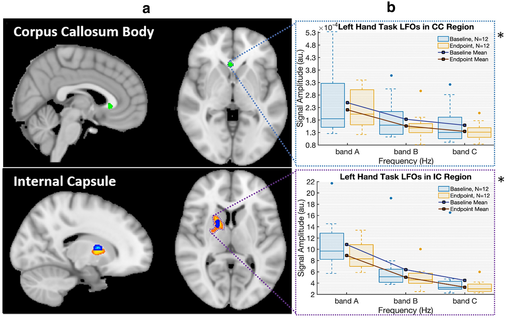
Read the figureA · The brain regions examined.B · Before-and-after signal measurements, grouped by frequency band.
Open the full-resolution figure ↗These independent studies inform the training rationale. NST’s specific curriculum and outcomes belong to the NeuroMotion research program ↗.
FROM OBSERVATION TO PRACTICE
Give the lesson a place in your week.
The curriculum provides the sequence. Each module connects an acquisition session to an application session, with room to mirror, revisit, and refine.
Open the 208-week pathway ↗